What Muscles Attach Left Hip And Back / Abdominal Muscles Function Anatomy Diagram Body Maps. The psoas is the primary hip flexor, assisted by the iliacus. What movements does it control? Hip flexor muscles attach the hip joints to the top of the femur and the inside of the knee, allowing flexibility of the upper leg. It extends from the upper arm bone to the hip bone and joins the abdominal and pectoral muscles. An increase in pressure within a muscular compartment because of the.
The psoas muscle is a deep muscle that connects your spine to your leg.in fact, it's the only muscle that does so. Related posts of muscles of the lower back and hip diagram human anatomy full picture. Check out the pilates ebook available here to find out more about what exercises will help to strengthen the hip muscles. The pectineus, the adductors longus, brevis, and magnus, as well as the tensor fasciae latae are also involved in flexion. When these muscles become tight due to inadequate activity (such as from a sedentary lifestyle), they become shorter, and in turn, cause tension around the sacroiliac.
Hip Flexor Pain Or Iliopsoas Related Groin Pain from mk0hippainhelp9h8quy.kinstacdn.com These muscles include the gluteus maximus muscle (the largest muscle in the body) and the hamstrings group, which consists of the biceps femoris, semimembranosus, and semitendinosus muscles. Hip flexor muscles attach the hip joints to the top of the femur and the inside of the knee, allowing flexibility of the upper leg. And, also like strains, tendonitis frequently affects the same population—athletes who participate in cycling, swimming, running, and other sports that repeatedly stress the hip. Check out the pilates ebook available here to find out more about what exercises will help to strengthen the hip muscles. In the back of the torso, the latissimus dorsi is a large, rectangular muscle that extends from the lower back near the top of the pelvis to near the shoulder. Side bending the trunk straightening of the spine (standing straight) The medial muscles of the hip are involved in the adduction of the leg i.e. The quadratus lumborum is a low back muscle that connects the hip bone (iliac crest), lower back vertebrae (l1, l2, l3, l4) to the 12 th rib.
When these muscles get tight, as they often do, you may find that along with hip pain, your lower back hurts—but you can't figure out why.
Bringing the leg back towards the midline. They are responsible for the range of motion in the legs and hips. Tension and knots in the trapezius muscles often occur due to stress and poor posture. Its primary role is in arm movement,. The left and right hip bones meet anteriorly at the body's midline in a band of fibrocartilage known as the pubic symphysis (or symphysis pubis). Hip muscles which make up the adductors The hip joint is a ball and socket synovial joint, formed by an articulation between the pelvic acetabulum and the head of the femur. Human anatomy full picture 12 photos of the human anatomy full picture human anatomy full picture, human anatomy pictures for artists online, human anatomy pictures free, human anatomy pictures kidney, human anatomy pictures muscles, human muscles, human anatomy full picture, human anatomy pictures for. Hip tendonitis is inflammation of any of the hip tendons, or thick cords that attach muscles to bone. Related posts of muscles of the lower back and hip diagram human anatomy full picture. Fibromyalgia is a chronic pain condition that can affect any joint or muscle in your body, including your left hip bone and buttock muscles. The psoas is the primary hip flexor, assisted by the iliacus. Many muscles that move the trunk and legs, such as our abdominal muscles, attach to the hip bones.
Human anatomy full picture 12 photos of the human anatomy full picture human anatomy full picture, human anatomy pictures for artists online, human anatomy pictures free, human anatomy pictures kidney, human anatomy pictures muscles, human muscles, human anatomy full picture, human anatomy pictures for. They are responsible for the range of motion in the legs and hips. Check out the pilates ebook available here to find out more about what exercises will help to strengthen the hip muscles. When these muscles become tight due to inadequate activity (such as from a sedentary lifestyle), they become shorter, and in turn, cause tension around the sacroiliac. Tension and knots in the trapezius muscles often occur due to stress and poor posture.
Why Physical Therapy And Yoga Did Not Help Your Low Back Pain Caring Medical Florida from www.caringmedical.com These muscles, including the gluteus maximus and the hamstrings, extend the thigh at the hip in support of the body's weight and propulsion. These muscles include the gluteus maximus muscle (the largest muscle in the body) and the hamstrings group, which consists of the biceps femoris, semimembranosus, and semitendinosus muscles. The largest of them is the most superficial muscle, the gluteus maximus. The back the lower back area, known as the lumbar spine, is made up of. There are three layers of gluteal muscles on the posterior hips, just like there are three layers of muscles in the abdominal trunk. The muscle is often described as having a long head (the attachment from the ischium) and a short head (attached to the femur). If a strain occurs on the left side of the body, it may cause pain above the left hip. Fibromyalgia is a chronic pain condition that can affect any joint or muscle in your body, including your left hip bone and buttock muscles.
An increase in pressure within a muscular compartment because of the.
Human anatomy full picture 12 photos of the human anatomy full picture human anatomy full picture, human anatomy pictures for artists online, human anatomy pictures free, human anatomy pictures kidney, human anatomy pictures muscles, human muscles, human anatomy full picture, human anatomy pictures for. Many muscles contribute to these movements: Obturator externus also helps to adduct the leg. If these muscles are stiff and tight, often due to remaining. All of these muscles of the hips have tendons at the end where they meet the bone. The hip ligaments attach the thighbone to the pelvis for added stability to the hip joint. The posterior muscle group is made up of the muscles that extend (straighten) the thigh at the hip. Muscles located at the side of the hip, which include the gluteus medius, piriformis, and hip external rotator muscles contribute greatly to the well being of your lower back, as well as your posture. When these muscles become tight due to inadequate activity (such as from a sedentary lifestyle), they become shorter, and in turn, cause tension around the sacroiliac. Hip tendonitis is inflammation of any of the hip tendons, or thick cords that attach muscles to bone. Tension and knots in the trapezius muscles often occur due to stress and poor posture. Pelvic muscles that cross the hip joint and attach onto the thigh/leg muscles that cross the hip joint are usually thought of with respect to their open chain motion of the thigh relative to the pelvis at the hip joint. As such, you can also divide the musculature that moves the thigh at the hip joint into quadrants.
They are responsible for the range of motion in the legs and hips. Related posts of muscles of the lower back and hip diagram human anatomy full picture. If a strain occurs on the left side of the body, it may cause pain above the left hip. The hip joint is a ball and socket synovial joint, formed by an articulation between the pelvic acetabulum and the head of the femur. The way your hip flexors and lower back muscles attach to the pelvis makes them particularly prone to this:
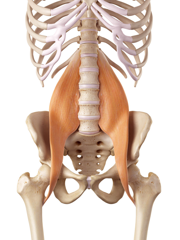
Psoas Constructive Rest Greenwood Physical Therapy from www.greenwoodpt.com It extends from the upper arm bone to the hip bone and joins the abdominal and pectoral muscles. The left and right hip bones meet anteriorly at the body's midline in a band of fibrocartilage known as the pubic symphysis (or symphysis pubis). The psoas is the primary hip flexor, assisted by the iliacus. It runs from your lower back through your pelvis, passing to the front of your hip where it attaches to the top of your femur, which is your thigh bone. The medial muscles of the hip are involved in the adduction of the leg i.e. As such, you can also divide the musculature that moves the thigh at the hip joint into quadrants. When these muscles become tight due to inadequate activity (such as from a sedentary lifestyle), they become shorter, and in turn, cause tension around the sacroiliac. The psoas muscle is a deep muscle that connects your spine to your leg.in fact, it's the only muscle that does so.
Reach forward with your right hand, pushing your right hip away from it.
Causes of muscle strains and stitches include: Gluteus maximus trigger point pain is felt toward the back of the hip and thigh near the hip joint, the base of the spine, and in the upper buttock going down alongside and into the gluteal fold. Doctors say that it's not uncommon for fibromyalgia to cause extreme hip discomfort. Many muscles contribute to these movements: Hip flexor muscles attach the hip joints to the top of the femur and the inside of the knee, allowing flexibility of the upper leg. The muscle is often described as having a long head (the attachment from the ischium) and a short head (attached to the femur). If your hip flexors are too tight (or too strong) in comparison to their opposing muscles, the glutes, then your lower back muscles are likely to end up tight too — and vice versa, if your lower back muscles are too tight in comparison to your abs. Fibromyalgia is a chronic pain condition that can affect any joint or muscle in your body, including your left hip bone and buttock muscles. As such, you can also divide the musculature that moves the thigh at the hip joint into quadrants. So can side stitches, a common and temporary athletic injury. These muscles include the adductors (adductor magnus, adductor longus, adductor brevis, pectineus, gracilis). The latissimus dorsi muscle is the widest and most powerful back muscle. Human anatomy full picture 12 photos of the human anatomy full picture human anatomy full picture, human anatomy pictures for artists online, human anatomy pictures free, human anatomy pictures kidney, human anatomy pictures muscles, human muscles, human anatomy full picture, human anatomy pictures for.